Educational activities are supported by the EURL-AP intranet
Micrographs collection
This collection, started in 2007, constitutes the largest repository of micrographs related to the detection of PAPs in feed. Through more than 800 high quality pictures taken by stereomicroscopy of light microscopy, the collection tends to illustrate the diversity of animal remains which can be found in feed. Typical illustrations of fish, farmed mammals, sea mammals, birds, rodents remains are to be found. Each picture contains separated information on the animal origin, the microscopic techniques used (bright field, polarized, DIC, phase contrast…), the embedding agent used, the staining, the magnification, etc. Every micrograph possesses a scale bar and an encrypted EURL-AP logo.

Periodic updates are realised at least twice a year and are announced by the intranet alert system. Content of the collection evolves with present issues usually raised by the NRL network during the annual workshop. Enrichment of the collection is supported by the large EURL-AP sample bank.
This collection is also accessible to IAG members (either public institution or private companies) on simple request. Searchers having access to the collection are free to use the pictures for publication provided they use a courtesy mention.
Protocols
Aside EU regulation, the next protocols developed by the EURL-AP can be found on the intranet:
- Preparation of permanent slides with a photopolymer
- Submission of samples to the EURL-AP for official counter analysis
- Conditions of image submission to the EURL-AP Micrograph Collection
- Test protocols for the improvement of the official light microscopic method according Annex VI of European regulation EC/152/2009
These protocols are restricted to the NRL members.
Training notes
Most of the training lectures are to be found on the EURL-AP intranet. Within a dedicated menu, NRL members can consult on-line or download the PDF formatted versions constituting the various training notes. This possibility is only granted to NRLs which are free to use the notes within the scope of their national training programs.
Tips and tricks
Special sheets are also likely to be found on the intranet in order to illustrate the best possible implementation of the methods for the detection of PAPs in feed. Up to now an extended leaflet covering all useful tips and tricks has been delivered to all NRL jointly with distribution of the DVD realised on the correct application of the official method.
Video sequences
By end of 2009, the EURL-AP team realised a DVD illustrating the best implementation of the official method. The video sequences illustrate different phases of preparation before final microscopic observation :
- Sample preparation and sedimentation process
- Sediment staining by Alizarin Red
- Sieving and slide mounting
- Some bad example that should be avoid
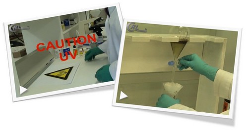
The intention of the video sequence which was offered to the NRLs in between December 2009 and February 2010 was to standardize as much as possible the sample preparation by using the official method. In this respect it also illustrates the use of the photopolymer used for the permanent slides. Actually over the years the EURL-AP demonstrated the key issue of slide mounting for a correct detection of animal proteins and their estimation of adulteration levels.
Based on this first realisation, in 2011 a second DVD was prepared for the correct implementation of PCR detection of animal proteins. This new DVD presents in five sequences the tips and tricks required for successful real time PCR analyses. This DVD was diffused to the NRLs. It also serves as a training support for the EURL-AP PCR trainings.